Median nerve: carpal tunnel and pronator
The median nerve can be compressed in the carpal tunnel and as it passes through pronator teres. This page brings both sites together with their anatomical plate and references. ULNT1 is the neurodynamic test that loads it.
Carpal tunnel
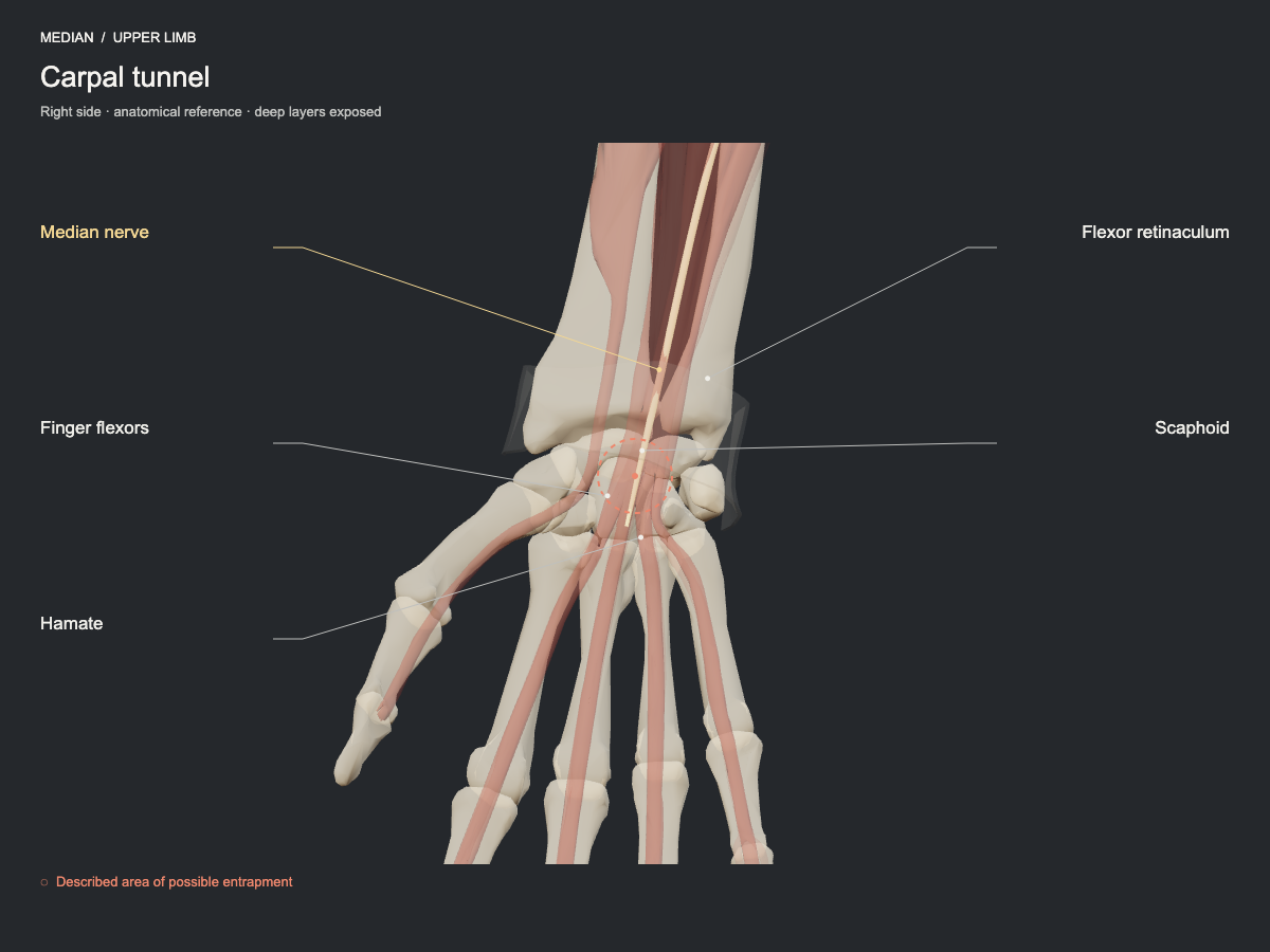
The median nerve crosses the tunnel alongside the flexor tendons. The carpal bones form the deep arch and the flexor retinaculum its roof. A reduction of the available space may compress the nerve; the palmar cutaneous branch runs superficial to the retinaculum and does not cross the tunnel.
- Frequency
- Common, well-established site for this nerve.
- Type of evidence
- Anatomical and clinical review
References
- Wu WT, Chang KV, Hsu YC, Tsai YY, Mezian K, Ricci V, Özçakar L. Ultrasound Imaging and Guidance for Distal Peripheral Nerve Pathologies at the Wrist/Hand. Diagnostics (Basel) 2023. PubMed 37296780 · DOI
Pronator teres region
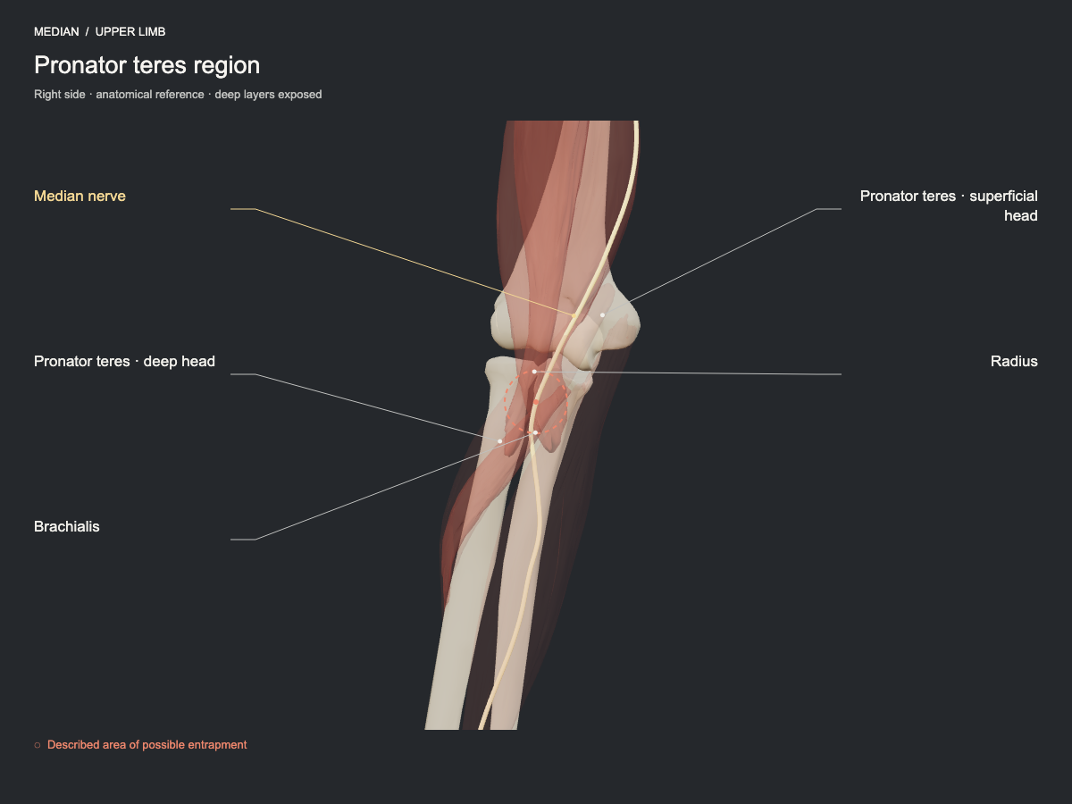
In the proximal forearm, the median nerve passes between the heads of pronator teres. Compression by the bicipital aponeurosis and by the arch of flexor digitorum superficialis is also described. These are proximal sites that must be differentiated from the carpal tunnel.
- Frequency
- Described site, less commonly seen.
- Type of evidence
- Anatomical and clinical review
References
- Choi SJ, Ahn JH, Ryu DS, Kang CH, Jung SM, Park MS, Shin DR. Ultrasonography for nerve compression syndromes of the upper extremity. Ultrasonography 2015. PubMed 25682987 · DOI
Tests that load this nerve
Mobilisation techniques
Median nerve · slider
As the elbow extends (the proximal bed lengthens), the wrist moves into palmar flexion (the distal bed shortens). On the way back the reverse happens.
Median nerve · tensioner
The elbow extends and the wrist moves into dorsiflexion at the same time: both ends lengthen the bed.
Joint parameters and animation in the techniques lab · Student plan €29.99/year
Evidence for the techniques
- Coppieters MW, Butler DS. Man Ther. 2008;13(3):213-21. PubMed 17398140 · DOI