Pronator teres syndrome: median nerve anatomy
In the proximal forearm the median nerve crosses several structures that can narrow its path: the bicipital aponeurosis, the two heads of pronator teres and the arch of flexor digitorum superficialis. This page places them on the 3D model and on the ultrasound image.
Anatomy of the passage
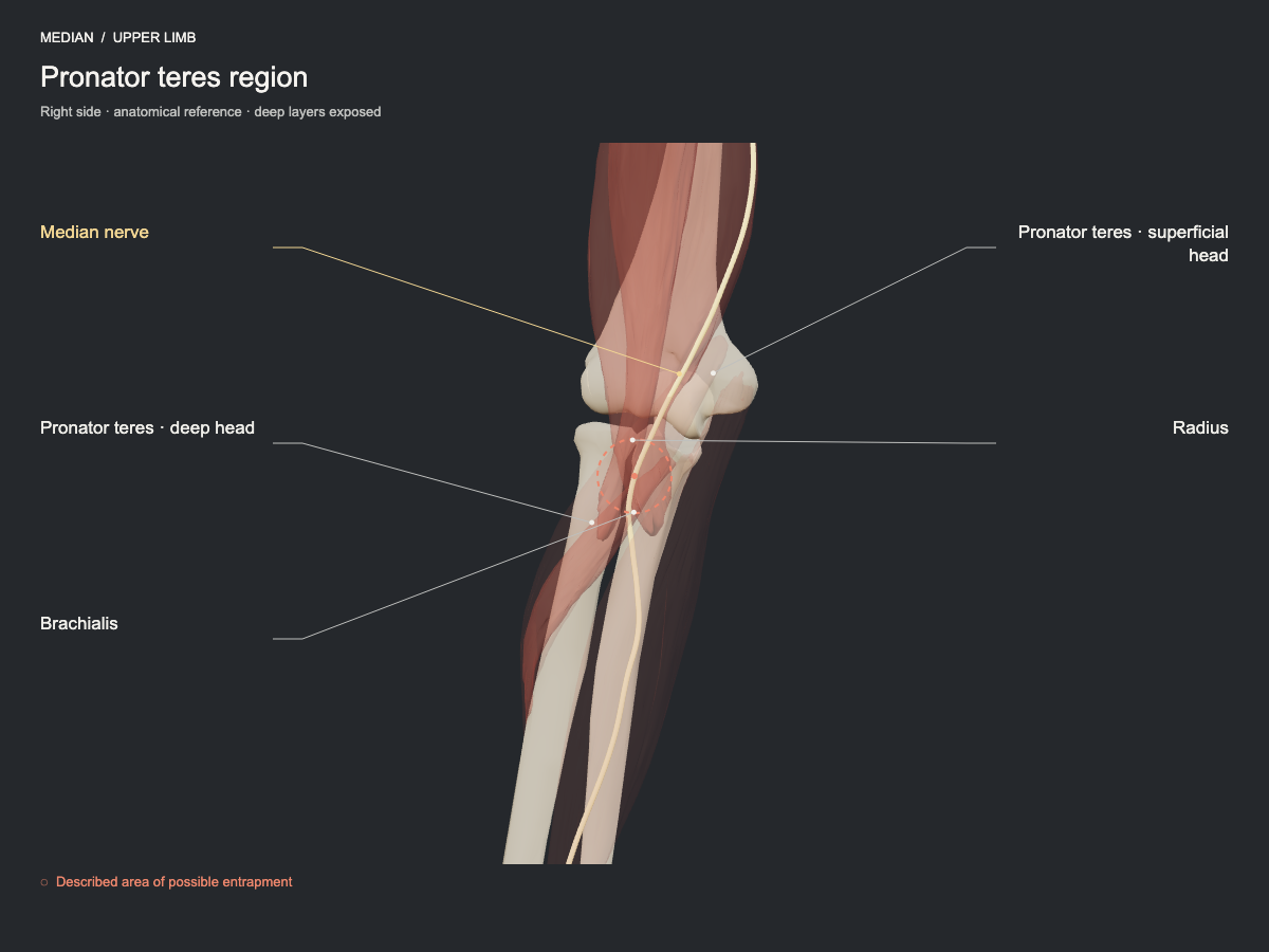
In the proximal forearm, the median nerve passes between the heads of pronator teres. Compression by the bicipital aponeurosis and by the arch of flexor digitorum superficialis is also described. These are proximal sites that must be differentiated from the carpal tunnel.
- Nerve
- Median nerve: carpal tunnel and pronator
- Frequency
- Described site, less commonly seen.
- Type of evidence
- Anatomical and clinical review
References
- Choi SJ, Ahn JH, Ryu DS, Kang CH, Jung SM, Park MS, Shin DR. Ultrasonography for nerve compression syndromes of the upper extremity. Ultrasonography 2015. PubMed 25682987 · DOI
Ultrasound landmarks
On the anterior aspect of the proximal forearm, the nerve passes between the superficial and deep heads of pronator teres and then beneath the arch of flexor digitorum superficialis. The brachial artery and its branches lie close by.
Tests that load this nerve
Other sites of the same nerve
All entrapment sites
Get atlas news
We will write to you when the plans open and when there is new content.