Carpal tunnel: anatomy of median nerve compression
The carpal tunnel is the best-known compression site of the upper limb. This page shows in 3D how the median nerve shares a rigid space with the flexor tendons beneath the flexor retinaculum, which ultrasound landmarks locate it and which neurodynamic test loads it.
Anatomy of the passage
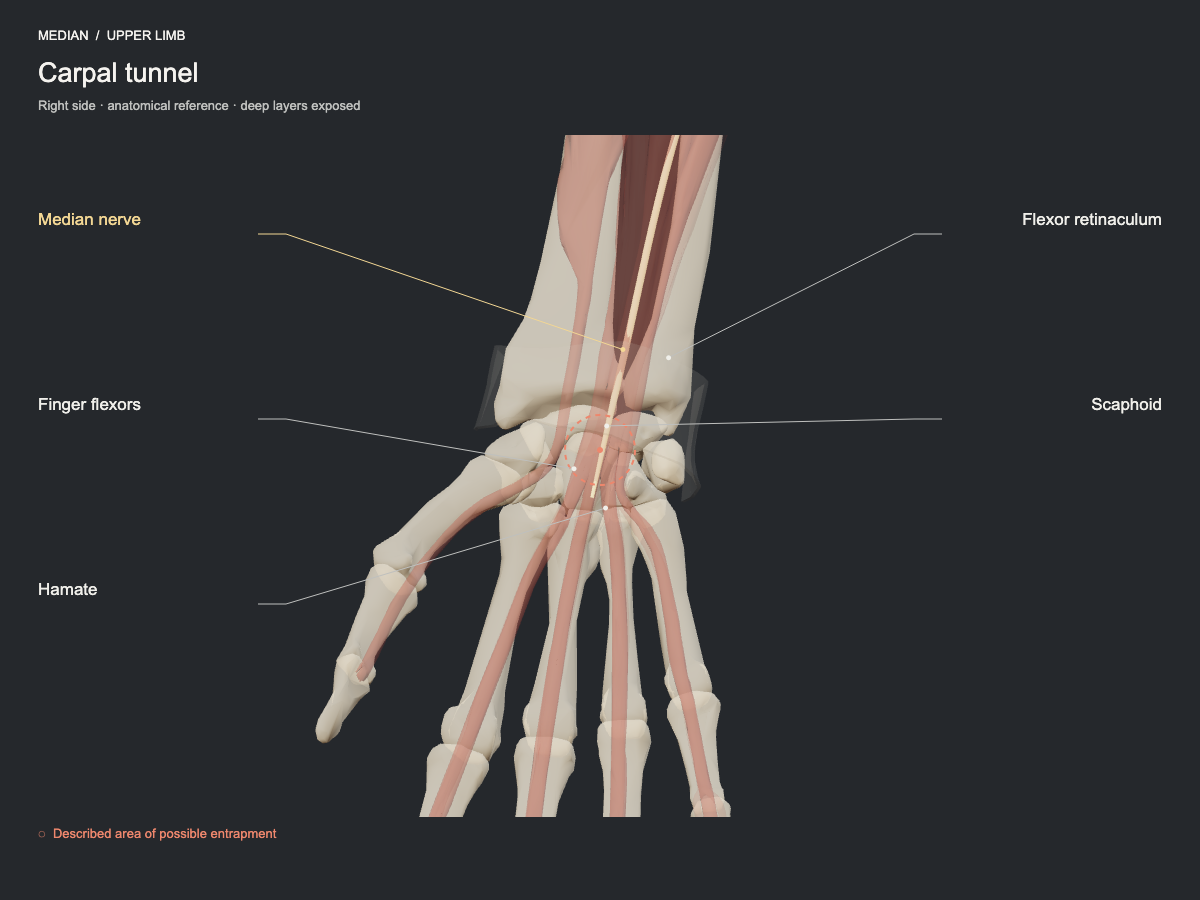
The median nerve crosses the tunnel alongside the flexor tendons. The carpal bones form the deep arch and the flexor retinaculum its roof. A reduction of the available space may compress the nerve; the palmar cutaneous branch runs superficial to the retinaculum and does not cross the tunnel.
- Nerve
- Median nerve: carpal tunnel and pronator
- Frequency
- Common, well-established site for this nerve.
- Type of evidence
- Anatomical and clinical review
References
- Wu WT, Chang KV, Hsu YC, Tsai YY, Mezian K, Ricci V, Özçakar L. Ultrasound Imaging and Guidance for Distal Peripheral Nerve Pathologies at the Wrist/Hand. Diagnostics (Basel) 2023. PubMed 37296780 · DOI
Ultrasound landmarks
As the probe moves from proximal to distal, the nerve passes beneath the flexor retinaculum alongside the flexor tendons; the pisiform and the scaphoid mark the proximal limit of the tunnel.
Tests that load this nerve
Other sites of the same nerve
All entrapment sites
Get atlas news
We will write to you when the plans open and when there is new content.