Tibial nerve: soleal arch and tarsal tunnel
The tibial nerve can be compressed beneath the proximal soleal arch and, more commonly, in the tarsal tunnel behind the medial malleolus. This page brings both sites together with their anatomical plate and references. The SLR and its ankle dorsiflexion variant are the neurodynamic tests that load it.
Proximal soleal arch
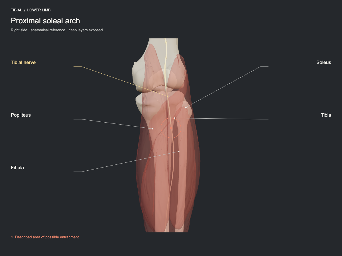
The tibial nerve descends through the popliteal fossa and passes deep to the proximal soleal arch. Compression by that fibrous arch or by space-occupying lesions in the region has been described. It is a proximal neuropathy less common than the distal site in the tarsal tunnel.
- Frequency
- Described site, less commonly seen.
- Type of evidence
- Narrative review
References
- Drăghici NC, Văcăraș V, Bolchis R, Bashimov A, Domnița DM, Iluț S, Popa LL, Lupescu TD, Mureșanu DF. Diagnostic Approach to Lower Limb Entrapment Neuropathies: A Narrative Literature Review. Diagnostics (Basel) 2023. PubMed 37958280 · DOI
Tarsal tunnel
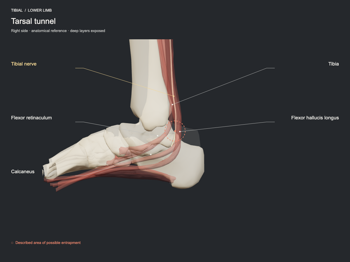
Behind the medial malleolus, the tibial nerve and its branches cross an osteofibrous space covered by the flexor retinaculum. Neighbouring tendons, ganglion cysts, fibrosis or anatomical variations may reduce the space. Involvement of the plantar branches explains why the symptoms are not identical in every case.
- Frequency
- Described site, less commonly seen.
- Type of evidence
- Anatomical and clinical review
References
- Bojovic M, Dimitrijevic S, Olory BCR, Eirale C, AlSeyrafi O, AlBaker AA, Krivokapic B, Jeremic D, DHooghe P. Overview of nerve entrapment syndromes in the foot and ankle. Int Orthop 2025. PubMed 40042611 · DOI