Soleal arch: tibial nerve entrapment
After the popliteal fossa, the tibial nerve and the posterior tibial vessels pass deep to the tendinous arch of soleus. It is an uncommon but described entrapment site. This page shows it on the 3D model and on the ultrasound image.
Anatomy of the passage
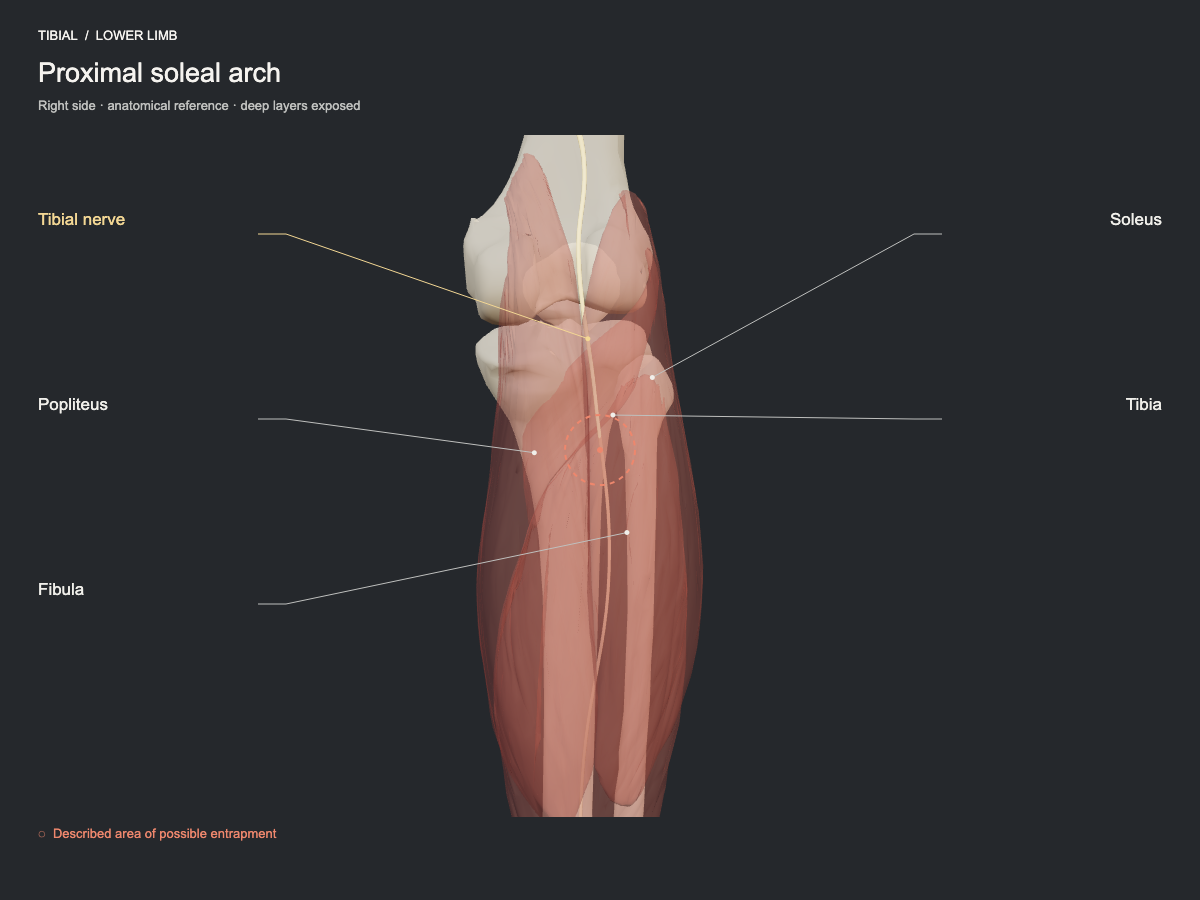
The tibial nerve descends through the popliteal fossa and passes deep to the proximal soleal arch. Compression by that fibrous arch or by space-occupying lesions in the region has been described. It is a proximal neuropathy less common than the distal site in the tarsal tunnel.
- Nerve
- Tibial nerve: soleal arch and tarsal tunnel
- Frequency
- Described site, less commonly seen.
- Type of evidence
- Narrative review
References
- Drăghici NC, Văcăraș V, Bolchis R, Bashimov A, Domnița DM, Iluț S, Popa LL, Lupescu TD, Mureșanu DF. Diagnostic Approach to Lower Limb Entrapment Neuropathies: A Narrative Literature Review. Diagnostics (Basel) 2023. PubMed 37958280 · DOI
Ultrasound landmarks
In the proximal posterior leg, the tibial nerve and the popliteal vessels pass beneath the tendinous arch of soleus, deep to the heads of gastrocnemius.
Tests that load this nerve
Other sites of the same nerve
All entrapment sites
Get atlas news
We will write to you when the plans open and when there is new content.