Tarsal tunnel: anatomy of tibial nerve compression
Behind the medial malleolus, the tibial nerve and its plantar branches pass through the tarsal tunnel beneath the flexor retinaculum, alongside the tendons and posterior tibial vessels. This page shows that space in 3D and the SLR with dorsiflexion that loads it.
Anatomy of the passage
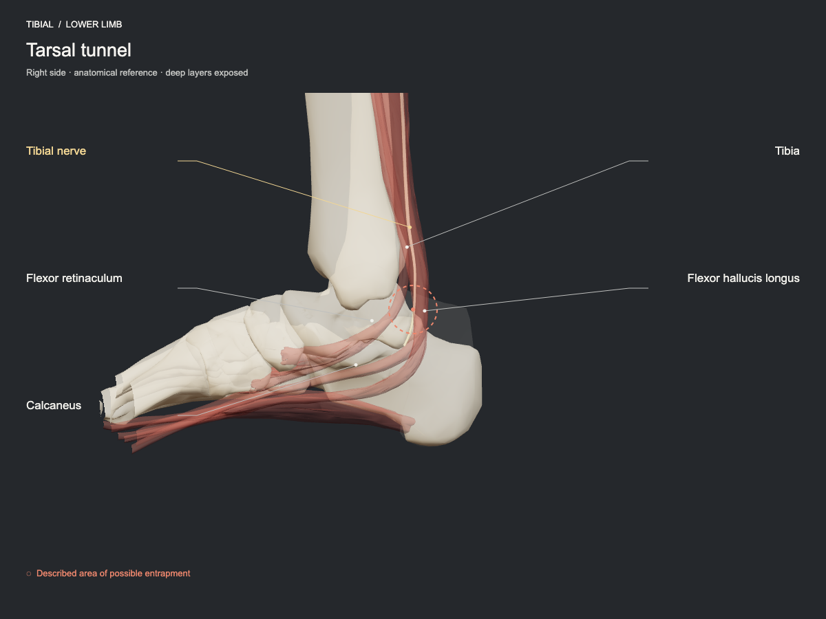
Behind the medial malleolus, the tibial nerve and its branches cross an osteofibrous space covered by the flexor retinaculum. Neighbouring tendons, ganglion cysts, fibrosis or anatomical variations may reduce the space. Involvement of the plantar branches explains why the symptoms are not identical in every case.
- Nerve
- Tibial nerve: soleal arch and tarsal tunnel
- Frequency
- Described site, less commonly seen.
- Type of evidence
- Anatomical and clinical review
References
- Bojovic M, Dimitrijevic S, Olory BCR, Eirale C, AlSeyrafi O, AlBaker AA, Krivokapic B, Jeremic D, DHooghe P. Overview of nerve entrapment syndromes in the foot and ankle. Int Orthop 2025. PubMed 40042611 · DOI
Ultrasound landmarks
Behind the medial malleolus, the nerve runs beneath the flexor retinaculum with the tendons of tibialis posterior, flexor digitorum longus and flexor hallucis longus, and divides into the medial and lateral plantar nerves. The posterior tibial artery and veins accompany the nerve inside the tunnel.
Tests that load this nerve
Other sites of the same nerve
All entrapment sites
Get atlas news
We will write to you when the plans open and when there is new content.