Sural nerve behind the lateral malleolus
The sural nerve is superficial behind the lateral malleolus, between the Achilles tendon and the fibular tendons. Trauma, scars or footwear can compress it. This page shows its course in 3D and the SLR with dorsiflexion that loads the tibial and sural pathway.
Anatomy of the passage
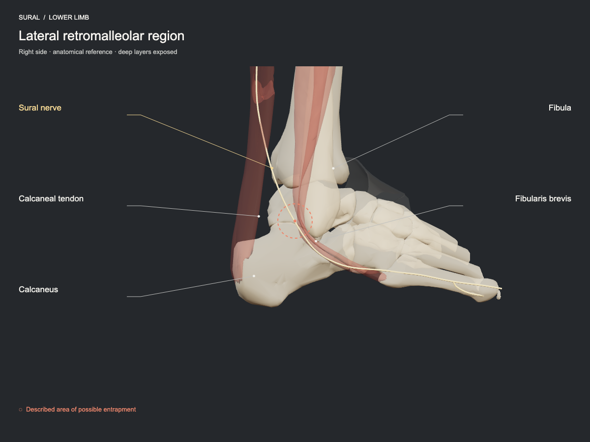
The sural nerve runs superficially behind the lateral malleolus, close to the calcaneal tendon and the fibularis tendons. Trauma, scars or external compression may affect it. It is sensory: its territory includes the lateral border of the foot, with variability between people.
- Nerve
- Sural nerve: lateral retromalleolar region
- Frequency
- Described site, less commonly seen.
- Type of evidence
- Anatomical and clinical review
References
- Bojovic M, Dimitrijevic S, Olory BCR, Eirale C, AlSeyrafi O, AlBaker AA, Krivokapic B, Jeremic D, DHooghe P. Overview of nerve entrapment syndromes in the foot and ankle. Int Orthop 2025. PubMed 40042611 · DOI
Ultrasound landmarks
Behind the lateral malleolus, the sural nerve runs subcutaneously with the small saphenous vein, superficial to the fibular tendons and lateral to the calcaneal tendon.
Tests that load this nerve
All entrapment sites
Get atlas news
We will write to you when the plans open and when there is new content.