Obturator canal: obturator nerve compression
The obturator nerve leaves the pelvis through the obturator canal, at the top of the obturator foramen, alongside the obturator vessels. Hernias, cysts or pelvic masses can compress it in that passage. This page shows it in 3D and the test that loads it.
Anatomy of the passage
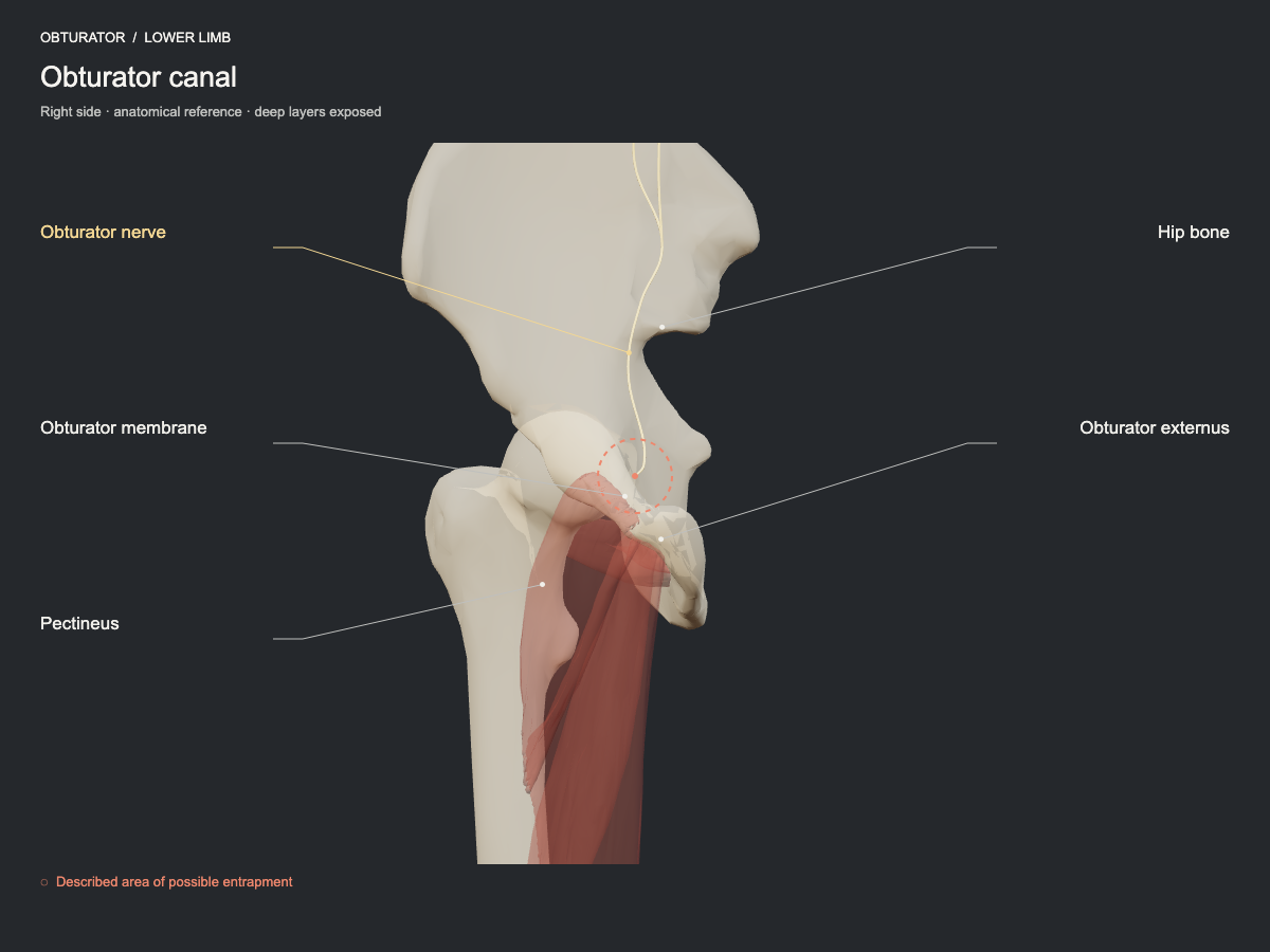
The obturator nerve leaves the pelvis through the canal in the upper part of the obturator foramen. Pelvic lesions, hernias, cysts or other masses may compress it. The neuropathy is uncommon; the cause and the level must be investigated individually.
- Nerve
- Obturator nerve: obturator canal and adductor planes
- Frequency
- Described site, less commonly seen.
- Type of evidence
- Review of clinical studies
References
- Scharfetter S, Wimmer F, Russe E, Schwaiger K, Pumberger P, Weitgasser L, Schaffler G, Wechselberger G. Endogenous Causes of Obturator Nerve Entrapment: Literature Review and Proposal of a Treatment Algorithm. J Clin Med 2025. PubMed 40142876 · DOI
- Kumar S, Mangi MD, Zadow S, Lim W. Nerve entrapment syndromes of the lower limb: a pictorial review. Insights Imaging 2023. PubMed 37782348 · DOI
Ultrasound landmarks
In the deep medial inguinal region, the obturator nerve leaves the obturator canal under the pectineus, over the obturator externus, accompanied by the obturator artery. Deep station.
Tests that load this nerve
Other sites of the same nerve
All entrapment sites
Get atlas news
We will write to you when the plans open and when there is new content.