Cubital tunnel: ulnar nerve compression at the elbow
At the elbow the ulnar nerve passes behind the medial epicondyle and enters between the heads of flexor carpi ulnaris. It is the second most common entrapment neuropathy of the upper limb. Its relations are shown here in 3D, together with the ULNT3 test that loads it.
Anatomy of the passage
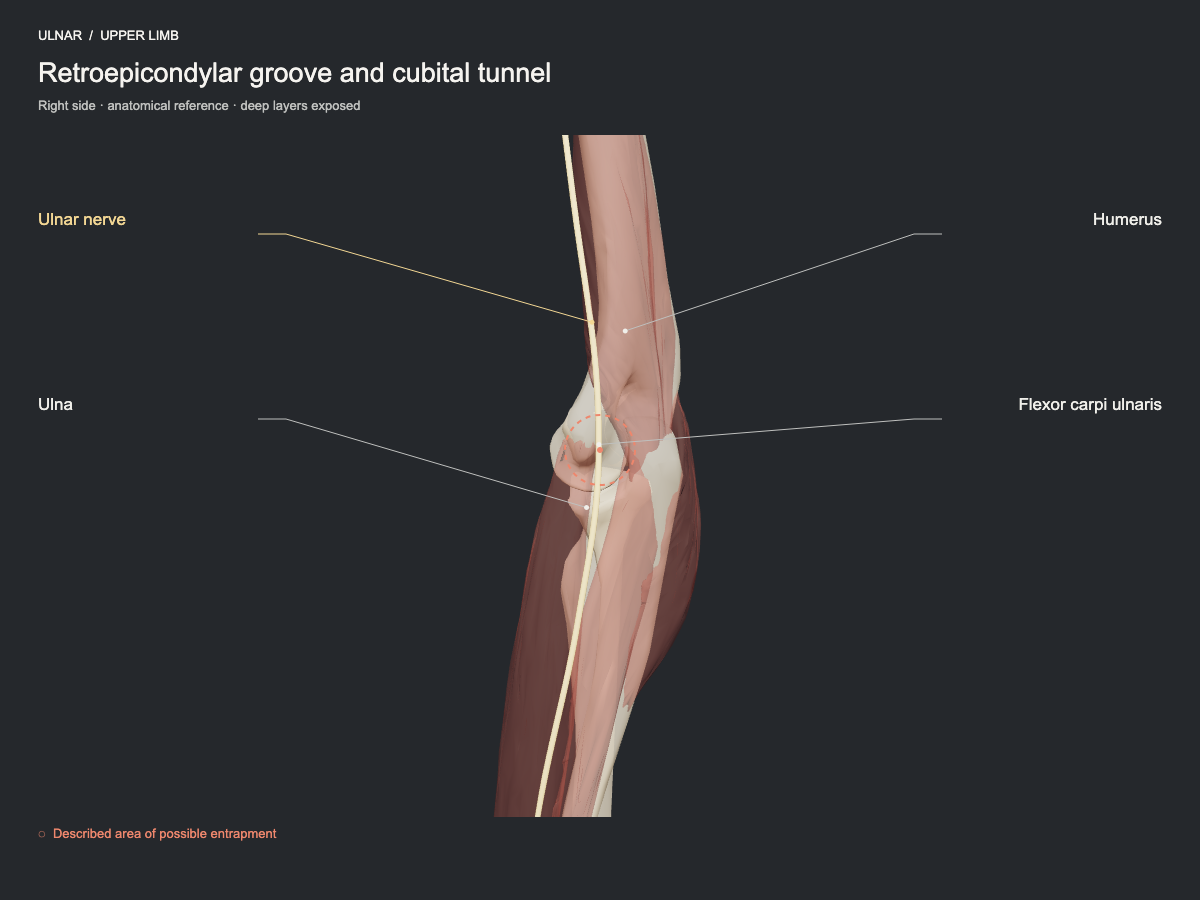
The ulnar nerve passes behind the medial epicondyle and enters between the heads of flexor carpi ulnaris. The fibrous roof of the tunnel, local bands and changes of space during elbow flexion may contribute to entrapment.
- Nerve
- Ulnar nerve: cubital tunnel and Guyon's canal
- Frequency
- Common, well-established site for this nerve.
- Type of evidence
- Anatomical and clinical review
References
- Choi SJ, Ahn JH, Ryu DS, Kang CH, Jung SM, Park MS, Shin DR. Ultrasonography for nerve compression syndromes of the upper extremity. Ultrasonography 2015. PubMed 25682987 · DOI
Ultrasound landmarks
In the retroepicondylar groove the nerve lies between the medial epicondyle and the olecranon; distally it enters between the heads of flexor carpi ulnaris. The roof of the tunnel (Osborne's ligament) is not segmented in this package.
Tests that load this nerve
Other sites of the same nerve
All entrapment sites
Get atlas news
We will write to you when the plans open and when there is new content.